


Understanding the anatomy of teeth is a vital aspect of maintaining good dental health. Our teeth play a crucial role in the process of digestion. As such, knowing the structure and functions of different tooth components can help us take better care of our oral health.
In this blog post, let’s delve into the fascinating world of tooth anatomy. Join us as we explore the various layers and parts of a tooth and their specific roles.
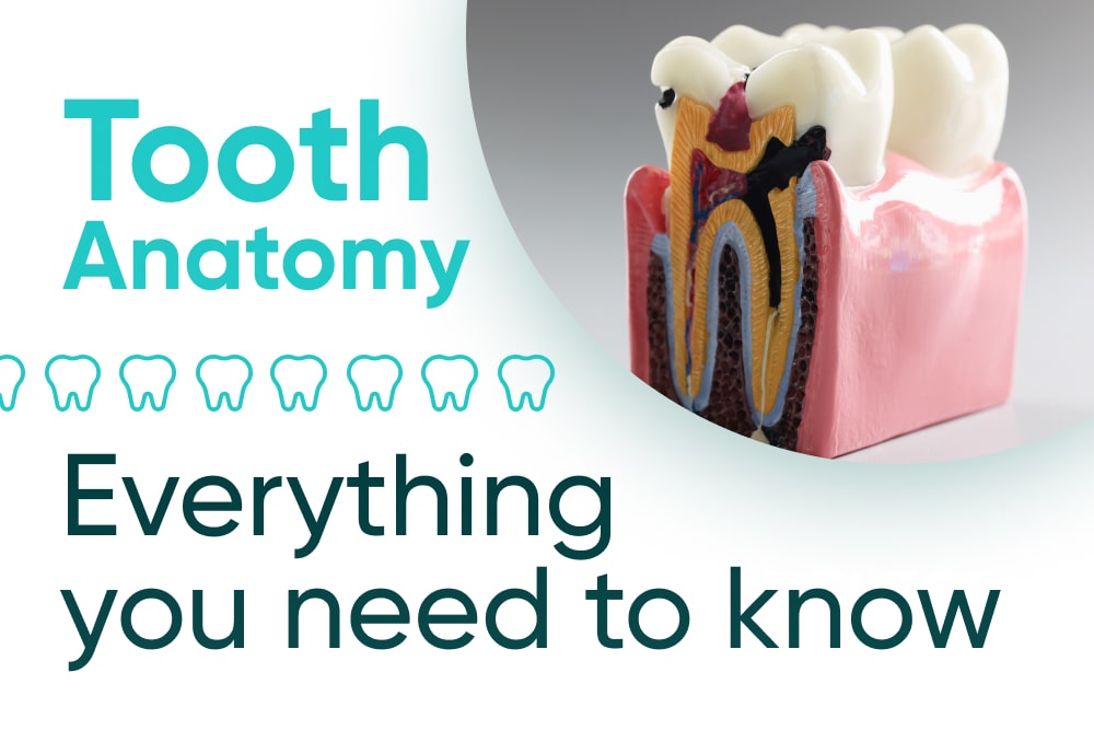
Anatomy of a tooth
Each tooth is a marvel of engineering, consisting of three main layers. Understanding the purpose of each layer is essential to grasp how teeth work together to facilitate chewing and speech.
1. Enamel: The protective shield
The outermost layer of a tooth is called the enamel. It acts as a remarkable shield that safeguards the tooth from various external threats.
The material for the enamel is an incredibly hard and mineral-rich substance. As such, it forms a robust barrier that covers the crown, which is the visible part of the tooth above the gum line.
The enamel plays a critical role in maintaining dental health. It does so by protecting the tooth from decay caused by harmful bacteria and acidic substances.
As we eat and drink, these harmful elements can attack the enamel. As such, it leads to the formation of cavities, eventually compromising the tooth’s integrity.
Thanks to its resilience and hardness, the enamel withstands the forces generated during chewing and biting. That’s why we can consume a wide variety of foods without damaging our teeth.
Without this protective layer, our teeth would be more susceptible to damage. Consequently, this can lead to discomfort and potential tooth loss.
However, despite its formidable strength, the enamel is not impervious to damage. The things we eat and do can gradually erode the enamel, such as:
- Poor oral hygiene
- Teeth grinding
- Excessive consumption of sugary or acidic foods
As a result, it is vital to adopt good oral hygiene practices, including regular brushing, flossing, and dental check-ups. This way, we can preserve the integrity of this protective shield and ensure the longevity of our teeth.
Plus, with the average price for dental services like cleaning falling between $200 and $1,200, it’s no wonder we should take proper care of our teeth.
2. Dentin: Supporting structure
Beneath the enamel lies the dentin. This is a softer but still resilient layer that forms the main bulk of the tooth. It plays a vital role in supporting the tooth’s structure and protecting its innermost core.
Dentin is a unique substance with a porous nature. It comprises tiny tubules that connect directly to the tooth’s nerves. As a result, dentin is highly sensitive to changes in temperature and pressure.
An intact enamel protects the dentin, which makes these sensations less noticeable. Conversely, a worn or damaged enamel exposes the dentin. This is why some people feel heightened sensitivity and discomfort when consuming hot or cold food or drinks.
Despite its sensitivity, dentin’s role as a supporting structure is crucial. As such, it acts as a shock absorber. This means that it helps distribute the forces generated during chewing and biting more evenly across the tooth.
This feature is important for the teeth located at the back of the mouth, like molars, which endure more substantial chewing forces.
With poor oral hygiene, dental cavities penetrate the enamel and reach the dentin. At this time, the cavities can progress more rapidly due to the latter’s softer nature. As a result, they become larger cavities and, in some cases, lead to infections in the pulp.
Maintaining good oral hygiene practices and addressing dental issues promptly is essential. As such, it helps you preserve the health and integrity of the dentin.
3. Pulp: The heart of the tooth
Deep within the tooth lies the pulp, occupying the central chamber or pulp chamber. Some describe it as the “heart” of the tooth.
The pulp is a delicate network of blood vessels, nerves, and connective tissues. As such, all of these play a pivotal role in keeping the tooth alive and healthy.
The pulp’s primary function is to supply essential nutrients and oxygen to the tooth. This way, it ensures each tooth’s vitality and overall health.
Additionally, the nerves within the pulp are responsible for providing sensations to the tooth. This allows us to perceive sensations like hot, cold, and pressure.
While the pulp is vital during tooth development, it can become less crucial after a tooth fully matures. In mature teeth, it’s the surrounding tissues and bone that provide support. Nonetheless, the pulp’s main role is still to continue supplying nutrients and maintaining the tooth’s sensitivity.
However, the pulp can become damaged or infected due to deep cavities, cracks, or trauma. This can lead to excruciating toothaches and other complications.
In such cases, a general dentistry expert may perform root canal treatment. The procedure entails removing the infected pulp and saving the tooth from extraction.
Preserving the health of the pulp is paramount for maintaining a healthy and functional tooth. As such, regular dental check-ups and early detection of dental issues can help prevent short and long-term damage.
Additional parts of the tooth
The tooth also has extended parts, such as the following:
1. Crown
2. Pulp chamber
Deep within the tooth, beneath the enamel and dentin layers, lies the pulp chamber. This chamber houses the vital pulp.
The pulp chamber extends from the crown all the way down to the tooth’s root. Its role is to protect the pulp, which nourishes and supplies the tooth with essential nutrients.
Additionally, the nerves in this chamber are responsible for transmitting sensations like pain, heat, and cold.
3. Root
Below the gum line, the tooth’s root is securely embedded within the jawbone. As such, it provides stability and support for the tooth.
Cementum, a thin layer, covers the roots, which helps anchor the tooth to the bone. The roots extend deep into the jaw, firmly holding the tooth in place.
This anchoring system is vital for withstanding the substantial forces generated during chewing and biting. Additionally, the roots serve as conduits. This means they allow blood vessels and nerves to travel from the jawbone to the pulp chamber.
4. Gum line
The gum line is the area where the gum tissue meets the tooth’s surface. This natural seal acts as a protective barrier. As such, it shields the sensitive tooth root from external stimuli, such as hot, cold, or acidic substances.
Healthy gums are essential for maintaining overall dental health. Basically, they help prevent bacteria from infiltrating the root and causing infections.
Regular brushing and flossing are crucial for keeping the gum line clean and free from harmful bacteria.
Tooth anatomy number
Medical professionals follow a standardized numbering system for tooth identification in dental anatomy. As a result, it streamlines communication and record-keeping during dental examinations and treatments.
This system facilitates efficient communication between dental professionals. With this, it’s easier to discuss specific teeth without any confusion.
The most commonly used tooth numbering system is the Universal Numbering System. In this method, each tooth is assigned a unique number.
Starting from the upper right side of the mouth and moving clockwise, the numbering goes as follows:
1. Upper right quadrant
Teeth are numbered from 1 to 8. It begins with the right upper third molar (wisdom tooth) as tooth number 1. Then, number 8 is the right upper central incisor.
2. Upper left quadrant
Here, teeth are numbered from 9 to 16. It starts with the left upper central incisor as tooth number 9. Then, it ends with the left upper third molar (wisdom tooth) as tooth number 16.
3. Lower left quadrant
With this, teeth are numbered from 17 to 24. The left lower third molar (wisdom tooth) is designated as tooth number 17. As such, the left lower central incisor is tooth number 24.
4. Lower right quadrant
Teeth are numbered from 25 to 32. It begins with the right lower central incisor as tooth number 25. Consequently, it concludes with the right lower third molar (wisdom tooth) as tooth number 32.
By using this numbering system, dentists can quickly and accurately refer to specific teeth during dental procedures. As a result, it ensures seamless communication and efficient patient care.
Human tooth anatomy: Permanent dentition
Individuals who have a full set of teeth should have 32 teeth. Each tooth falls under one of these categories based on its position:
1. Canine tooth anatomy
You can find canine teeth or cuspids at the corners of the mouth. You may know them through their pointed shape, hence the moniker.
Canines play a crucial role in tearing and ripping food. Moreover, they’re particularly helpful for consuming meat and raw vegetables. As such, these teeth have long, robust roots that provide stability during biting and chewing.
Canines typically emerge during childhood, usually around the ages of nine to 12 years.
Proper alignment of canines ensures an aesthetically pleasing smile and an efficient chewing process. With proper dental care, whether through orthodontics, we can preserve the functionality and appearance of these important teeth.
2. Incisor tooth anatomy
Incisors, commonly known as front teeth, are the first to appear in the mouth. Front tooth anatomy shows that they have a flat and chisel-shaped edge. As such, this makes them perfect for cutting and biting food.
Four incisors are present in the front of the upper jaw and four in the front of the lower jaw. These teeth play a crucial role in the initial stages of food consumption. With them, you can slice through fruits, vegetables, and soft foods efficiently.
Incisors have relatively small roots, securely anchoring them to the jawbone for stability. These front teeth typically emerge in children between the ages of six and eight years.
3. Premolar tooth anatomy
Premolars or bicuspids are versatile teeth located behind the canines. They have a flatter and broader surface. As such, they’re responsible for tearing, crushing, and grinding food.
There are two premolars in each quadrant of the mouth. It’s crucial to note that they prepare food for further digestion by the molars.
Premolars have one or two roots, depending on their location, providing stability for their chewing function. Typically, the first premolars emerge around the age of 10 to 12 years, followed by the second premolars.
4. Molar tooth anatomy
Molars are large and robust teeth located at the back of the mouth. They’re primarily responsible for grinding and chewing food into smaller, digestible particles.
They have extensive flat surfaces with multiple raised points called cusps. Each quadrant of the mouth contains three molars:
- First molars – the third to the last tooth on each side of the mouth
- Second molars – the second to the last tooth on each side of the mouth
- Third molars – the last tooth at the back of the mouth; also known as wisdom teeth
Molars have three roots that securely anchor them to the jawbone, providing stability during the intense chewing process.
The first molars appear around six years of age, followed by the second molars around 12 years old.
5. Wisdom tooth anatomy
Wisdom teeth, also known as third molars, are the last set of molars to emerge in the mouth. They usually come out during late adolescence or early adulthood.
You can find your wisdom teeth at the back of the mouth. Additionally, there are four wisdom teeth in total. There’s one on each side of both the upper and lower jaws.
People often face challenges during wisdom teeth eruption. One such complication is impaction.
An impacted wisdom tooth can cause pain, swelling, and gum inflammation. Moreover, it can lead to infections and misalignment of neighboring teeth.
To prevent complications and maintain oral health, dentists often recommend wisdom tooth extraction when necessary. Regular dental evaluations help manage wisdom teeth effectively, promoting overall oral health and preventing potential problems.
Conclusion
With all these layers, it’s no wonder that an ache in a single tooth can disrupt your daily life. To learn about your teeth’s positions and how it affects you, you can check a tooth anatomy chart.
As such, having a comprehensive knowledge of tooth anatomy empowers us to make informed decisions about our dental health.
Proper oral hygiene practices and a balanced diet contribute to maintaining strong teeth throughout our lives. Additionally, going for regular check-ups at a trusted dental clinic like NoHo Family Dental can help preserve your smile.
Visit us now and enjoy a lifetime of optimal oral health starting today.